SPECTRALIS OCT

Benefits of a SPECTRALIS examination
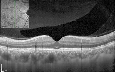
A SPECTRALIS® OCT examination provides a series of high-resolution digital images of the back of your eye, which give information about the condition of the retinal layers and the optic nerve head. These images provide the microscopic detail needed to help identify early signs of potential sight threatening diseases, such as diabetic retinopathy, glaucoma, and age-related macular degeneration. It is important to understand that early detection can help preserve sight.
If you are diagnosed with an eye disease, managing your condition will likely require follow-up visits, during which the doctor may take more images of your eye to make sure the disease is not progressing. SPECTRALIS offers comprehensive examination, as your eye care professional can conduct multiple tests with just one instrument. This, combined with the instrument’s ability to scan the exact same part of your eye in every follow-up-examination provides the assurance that small changes in the retina will be detected and managed for the best possible result. It is important to do your part and keep all follow-up appointments.
The examination process

You will be seated directly in front of the instrument and rest your chin on the adjustable chin rest. Separated by a protective breath shield, the examiner will be seated opposite of you on the other side of the instrument. You will look directly into the camera, at a fixation point and follow the instructions of the examiner. You can blink normally, and the examination will only take a few minutes.
When the examination begins, a small cross will appear in the instrument, which you will be asked to fixate. The examiner will then slowly move the instrument in your direction, without touching you. Within a safe distance to your eye, a precise image is captured of the various layers of your retina or optic nerve head. A harmless beam of light will scan the most important structures.
After the examination
The examination is completely painless and contact-free. Your vision will not be impaired. Unless your eye care professional has dilated your pupil to conduct the OCT test or another examination, you will be able to drive as usual.